Respiratory symptoms rarely point to one diagnosis or one test. By the end, you will know how a complete evaluation is assembled, which investigations fit common symptoms, when breathing problems require emergency care, and how to judge whether a chest hospital can manage the full path from diagnosis to follow-up.
Key takeaways
- Match spirometry, imaging or bronchoscopy to the symptom and clinical question.
- Seek emergency care for severe breathlessness, blue lips, confusion or coughing blood.
- Ask how test results change treatment, follow-up timing and referral decisions.
- Compare hospitals by respiratory specialists, intensive care, diagnostic tests and thoracic surgery access.
What does a complete respiratory evaluation include?
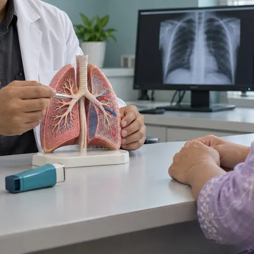
No single test proves a complete respiratory diagnosis. A complete respiratory evaluation builds evidence in sequence, then a specialist interprets the findings together.
1. Start with symptom history: cough, wheeze, breathlessness, chest pain, fever, weight loss and coughing blood. Record smoking, occupational dust or chemical exposure, biomass-smoke exposure, current medicines and inhaler use.
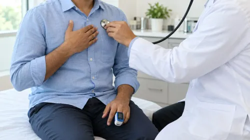
2. Check the respiratory rate, pulse, temperature, chest examination and oxygen saturation. Pulse oximetry detects low oxygen but does not measure carbon-dioxide retention. Motion, poor circulation, nail products and abnormal haemoglobin can distort the reading.
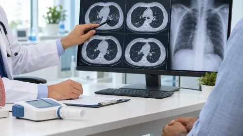
3. Use a chest X-ray or CT when infection, obstruction, cancer, scarring or another structural problem is suspected. Blood tests and sputum testing add evidence for inflammation, infection or tuberculosis.
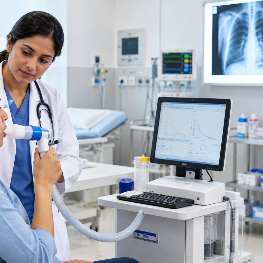
4. A pulmonary function test, especially spirometry, is central when airflow obstruction is suspected. Results must be technically acceptable and interpreted against appropriate reference equations. In adults with the right clinical picture, a post-bronchodilator FEV1/FVC ratio below 0.70 for COPD diagnosis confirms persistent obstruction; symptoms alone do not.
5. Normal spirometry at one visit does not rule out asthma. Bronchodilator reversibility, serial peak-flow measurements or bronchial provocation testing can demonstrate variable airflow limitation.
Severe illness or suspected ventilatory failure requires an arterial or venous blood-gas test, which measures oxygen, carbon dioxide and acidity more directly.
Which test fits your symptom, and when is it an emergency?
The right first investigation depends on the symptom, its duration and your clinical stability; no fixed checklist diagnoses every respiratory illness. Persistent cough tests usually begin with a chest X-ray, spirometry, medication and smoking or exposure review. Add sputum testing when infection is suspected.
| Symptom | Sensible first investigations | What changes the next step |
|---|---|---|
| Persistent cough | Chest X-ray, spirometry, exposure and medication review | Sputum testing for suspected infection; CT or bronchoscopy for persistent abnormalities |
| Breathlessness and wheezing evaluation | Oxygen saturation, chest examination and spirometry | Imaging or arterial/venous blood-gas analysis if severe, unexplained or associated with ventilatory failure |
| Coughing blood | Prompt imaging and blood tests | The amount, recurrence and your stability determine observation, CT, bronchoscopy or admission |
| Chest pain | Urgent assessment for lung, pleural, cardiac and vascular causes | Emergency testing when pain is sudden, severe or accompanied by breathlessness |
| Repeated pneumonia investigation | Review prior images, microbiology and immune or aspiration risks | CT, bronchoscopy or tissue sampling for recurrence, weight loss or an abnormal X-ray |
Seek emergency care for severe breathlessness at rest, blue lips, confusion, sudden chest pain, coughing a large amount of blood, rapidly falling oxygen saturation, or a reading around 90% or lower. Do not wait for a routine appointment.
A pulse oximeter does not measure carbon dioxide and readings can be distorted by motion, poor circulation, nail products or abnormal haemoglobin; interpret the number alongside your condition.
How do patients move from diagnosis to treatment and follow-up?
After first consultation, a chest specialist reviews symptoms, examination, oxygenation, lung-function, imaging, blood and sputum results together. The diagnosis may be asthma, COPD, pneumonia, tuberculosis, bronchiectasis, pleural effusion, pneumothorax, interstitial lung disease, sleep apnoea or lung cancer; test review also shows when further sampling or imaging is needed.
- Confirm the working diagnosis, severity and immediate risks.
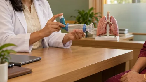
- Treat the cause: correct inhaler technique; prescribe antibiotics or anti-tuberculosis therapy when indicated; create an airway-clearance plan; drain selected pleural effusions or pneumothoraces; use disease-specific medicines; support smoking cessation.
- At follow-up, reassess symptoms, oxygenation, lung function, imaging, treatment response and adverse effects.
- Refer for surgery or make a respiratory intensive care referral when disease, deterioration or test results require it.
Pulmonary rehabilitation is prescribed exercise, education and behaviour change, not generic fitness advice. Oxygen therapy raises oxygen levels; high-flow nasal oxygen, non-invasive ventilation and invasive mechanical ventilation solve different problems.
In acute COPD exacerbation with respiratory acidosis, commonly pH 7.35 or lower with raised PaCO2, non-invasive ventilation provides mask-based ventilatory assistance under close monitoring, not simply stronger oxygen. Worsening exhaustion, shock, refractory hypoxaemia or inability to protect the airway can require urgent intubation.
When do bronchoscopy, biopsy and lung surgery become necessary?
Bronchoscopy becomes necessary when imaging, sputum tests or symptoms leave an airway problem, infection or suspected cancer unresolved. It directly examines the trachea and bronchi, collects washings, brushings or biopsies, removes secretions or foreign bodies, and assesses selected obstructions.
Flexible versus rigid bronchoscopy is a choice based on the task, not a universal ranking. Flexible bronchoscopy usually passes through the nose or mouth after local anaesthetic and sedation; recovery is shorter, but bleeding, infection, low oxygen and lung injury are bronchoscopy biopsy risks.
Rigid bronchoscopy uses a larger tube under general anaesthesia, providing a wider working channel and stronger airway control for major obstructions, bleeding control or larger instruments. Follow the treating team’s instructions for fasting, medicines, anaesthesia and recovery.
- Use CT alongside bronchoscopy; the procedure does not replace CT or pulmonary function testing. A negative sample may miss cancer or infection when the lesion is difficult to reach.
- Plan lung cancer tissue diagnosis and staging together. Preserve tissue for histology, molecular alteration testing and PD-L1 testing, then review surgery, stereotactic or conventional radiotherapy, chemotherapy, immunotherapy and targeted treatment with a multidisciplinary team.
- Before resection, FEV1 and DLCO before lung surgery, plus exercise capacity, guide risk. Video-assisted or robot-assisted surgery suits selected tumours, not every tumour; open surgery or non-surgical treatment may be safer.
How are sleep, tuberculosis and screening investigations chosen?
Symptom-driven diagnosis is different from screening. Sleep apnoea diagnosis requires sleep testing, not symptoms alone. An apnoea-hypopnoea index of 5 or more with relevant symptoms or comorbidity, or 15 or more regardless of symptoms, is commonly used. Home sleep-apnoea testing suits selected adults without major complicating conditions.
For suspected pulmonary tuberculosis, use this investigation sequence:
- A chest X-ray can show lung abnormalities but cannot establish infectious TB or drug susceptibility.
- A pulmonary tuberculosis molecular test can detect Mycobacterium tuberculosis and some resistance markers.
- Link a positive result to culture, susceptibility testing and public-health treatment pathways.
Low-dose CT lung cancer screening is for high-risk people without symptoms, not a general lung check-up. US Preventive Services Task Force criteria are annual low-dose CT from age 50 to 80 for adults with at least a 20 pack-year history who currently smoke or quit within 15 years. Local programmes may use different eligibility rules.
If symptoms are present, arrange diagnostic assessment rather than screening.
How should you compare a chest hospital with a general hospital?
A practical chest hospital versus general hospital comparison asks whether respiratory expertise and key investigations are available in one pathway.
| Facility feature | Check before booking | Why it matters |
|---|---|---|
| Specialists | Pulmonologists, thoracic surgeons and respiratory therapists | Complex cases receive focused review |
| Testing | Pulmonary function testing, imaging, flexible and rigid bronchoscopy, and sleep testing | The next test matches the suspected condition |
| Support | Respiratory intensive care and emergency support | Deterioration does not require a separate transfer |
| Coordination | Medical treatment, drainage, biopsy, surgery, ventilation and follow-up | Fewer handoffs and repeated appointments |
In Trichy, use this checklist when assessing Galaxy Chest & Multi Specialty Hospital Pvt Ltd and ask which services suit your specific condition. Confirm whether difficult cases are reviewed jointly rather than passed between departments.
- Prior CT or X-ray images, radiology and pathology reports
- Medicine list with doses, inhalers and discharge summaries
- Sputum or blood-test results, previous treatment records and a symptom timeline
- Smoking, occupational or biomass exposure, exercise tolerance and emergency episodes
Ask which test comes first, its preparation, when results will be reviewed, and which findings would trigger surgery or intensive care referral.
Frequently asked questions
What does a complete respiratory evaluation include?
It combines your history and examination with tests such as oxygen measurement, chest imaging, blood tests, spirometry or other lung-function studies. A specialist interprets the results together rather than relying on one test.
Which respiratory symptoms require emergency care?
Seek urgent care for severe or rapidly worsening breathlessness, blue or grey lips, confusion, fainting, chest pain, or coughing up a large amount of blood.
When is bronchoscopy or a lung biopsy necessary?
Doctors consider bronchoscopy when they need to inspect the airways, collect samples or remove an obstruction. A biopsy is used when tissue is needed to identify cancer, infection or another lung disease.
How are tuberculosis, sleep disorders and screening investigations chosen?
The clinician selects tests from your symptoms, exposure history, examination and risk factors. Options can include sputum testing or chest imaging for tuberculosis, a sleep study for suspected sleep apnoea, and targeted screening rather than indiscriminate testing.
How should you compare a chest hospital with a general hospital?
Check whether the hospital offers respiratory specialist assessment, lung-function testing, advanced imaging, bronchoscopy, pathology, respiratory critical care and thoracic surgery with coordinated follow-up.