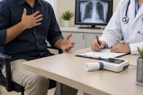
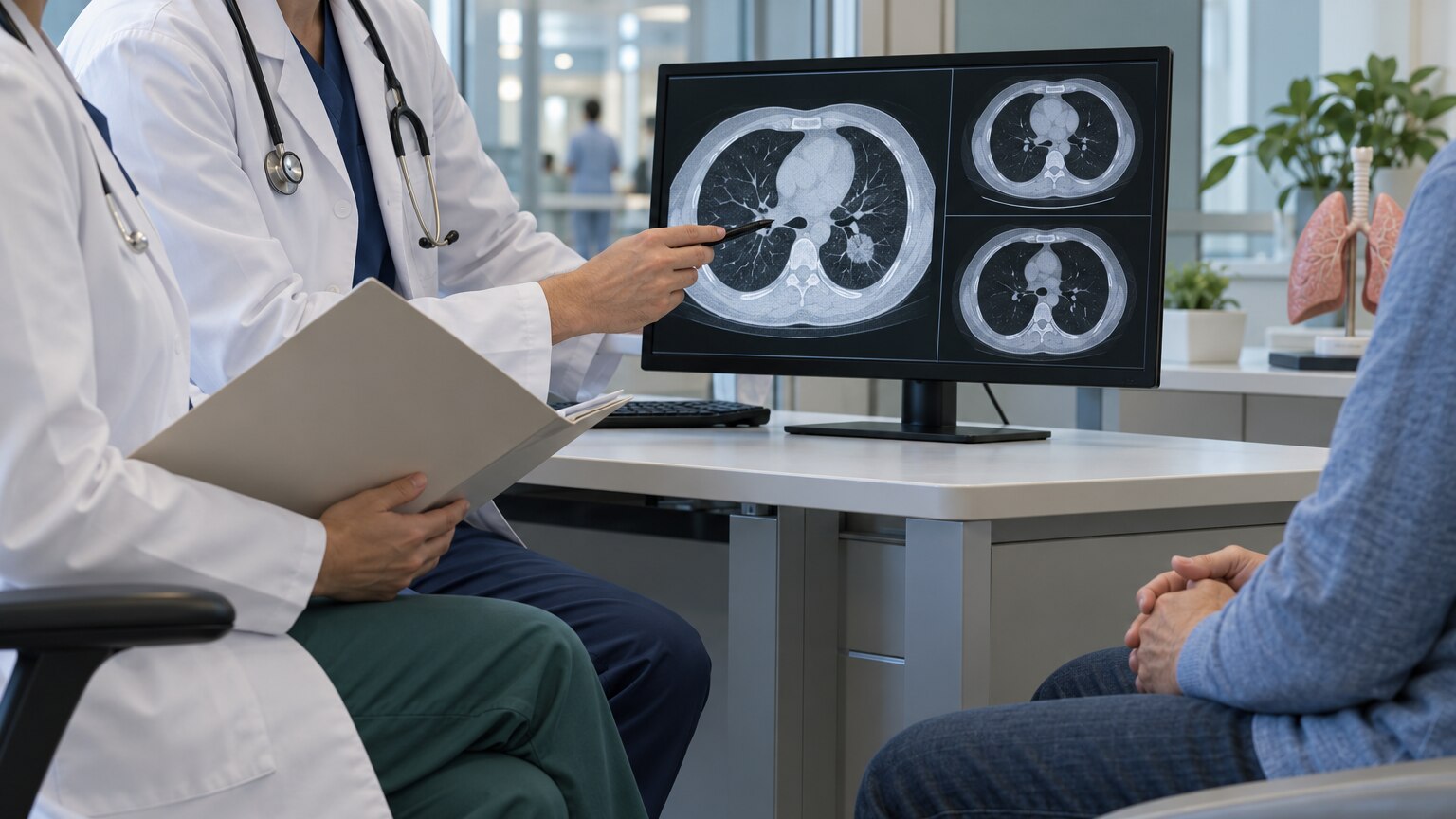
A suspicious lung symptom or scan finding does not answer three crucial questions: whether the problem is cancer, how far it has spread and which treatment is safest. By following the diagnostic pathway, checking the right tests and preparing for treatment decisions, you can ask more useful questions and recognise when respiratory symptoms require emergency care.
Key takeaways
- Seek urgent assessment for coughing blood, severe breathlessness or rapidly worsening chest pain.
- A suspicious scan is not a cancer diagnosis; tissue pathology usually confirms the tumour type.
- Ask how stage, molecular test results and fitness for surgery affect your treatment plan.
- Bring scan images, pathology slides and a complete medication list to multidisciplinary reviews.
When a lung symptom needs urgent assessment or emergency care
A persistent cough, coughing blood, unexplained weight loss or a non-resolving chest infection warrants prompt lung-cancer evaluation, especially with smoking history, occupational exposure, chest pain or new hoarseness. Book a clinician review for chest imaging and appropriate sputum, microbiology or tissue testing; do not assume tuberculosis or an infection explains symptoms indefinitely.
Tuberculosis and lung cancer can resemble each other on scans and can coexist, and improvement after antibiotics or tuberculosis treatment does not by itself exclude cancer.
| Situation | What to do | Why it matters |
|---|---|---|
| Persistent cough, blood-streaked sputum, unexplained weight loss or a non-resolving infiltrate | Arrange prompt respiratory assessment | Cancer, tuberculosis, infection and scarring can overlap |
| Breathlessness at rest, rapidly worsening breathing or inability to speak full sentences | Seek emergency respiratory care now | Low oxygen or airway compromise can deteriorate quickly |
| Large-volume or repeated coughing blood, blue lips, confusion, fainting or severe chest pain | Call emergency services or go to an emergency department | These signs can indicate major bleeding, respiratory failure or another life-threatening problem |
Do not drive yourself if you are severely breathless, confused or coughing significant blood. Emergency care stabilises breathing and bleeding first; oncology investigations follow once you are safe.
How screening and chest scans separate suspicion from diagnosis
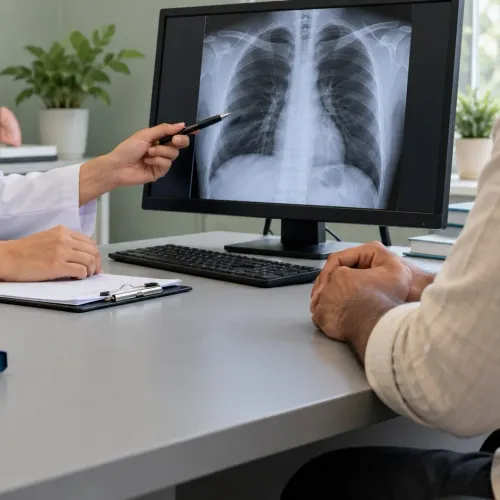
A scan can raise suspicion; a tissue sample establishes whether a nodule is cancer.
| Test | What it shows | When it applies |
|---|---|---|
| Low-dose CT | Thin-slice images with less radiation | Annual lung-cancer screening for eligible high-risk adults |
| Diagnostic CT | Detailed images, often with intravenous contrast | Characterising a nodule, mass, lymph nodes or complications |
| Chest X-ray | A quick, broad two-dimensional view | Initial assessment; it can miss small or hidden cancers |
| PET-CT | Areas with increased tracer uptake across the body | Mapping metabolically active disease for staging; uptake is not proof of cancer |
| Brain MRI | High-detail images of the brain | Suspected brain spread or staging protocols; PET-CT is less sensitive in the brain |
Adults aged 50–80 with a 20 pack-year smoking history who currently smoke or quit within 15 years should discuss annual low-dose CT. Occupational exposure to asbestos, silica, diesel exhaust or other carcinogens also warrants a risk review; chest X-ray is not an equivalent screening test.
For a nodule, clinicians compare older scans, measure size and growth, and assess its shape, location and smoking exposure. Follow-up CT, sputum or other microbiology, bronchoscopy, or needle biopsy can distinguish cancer from tuberculosis, infection, scarring or a benign growth. A temporary response to antibiotics does not rule out cancer.
Choosing a biopsy and understanding the pathology report
Choose the safest biopsy route by matching the lesion’s location, size, airway access, nearby blood vessels, lung function and need to sample lymph nodes. Preserve enough tissue for both diagnosis and biomarker testing.
| Option | Best suited for | Main trade-off |
|---|---|---|
| Bronchoscopy-guided biopsy | Lesions near airways | Smaller sample; sedation and bleeding risks |
| CT-guided percutaneous biopsy | Peripheral lung nodules | Pneumothorax risk |
| EBUS-TBNA | Mediastinal or hilar lymph nodes | A negative result may need confirmation |
| Surgical biopsy | Inaccessible or repeatedly nondiagnostic lesions | General anaesthesia and recovery |
- Which route gives the highest diagnostic yield with my lung function?
- Will you sample lymph nodes and send material for microbiology, including tuberculosis testing?
- How much tissue will be reserved for molecular tests and PD-L1 staining?
- What are the bleeding, pneumothorax, anaesthesia and hospital-stay risks?
The pathology report should identify the cancer type, such as adenocarcinoma, squamous-cell carcinoma or small-cell carcinoma, and state whether the sample is adequate. For non-small-cell lung cancer, testing commonly includes EGFR, ALK, ROS1, BRAF, KRAS, MET, RET and NTRK alterations.
The PD-L1 tumour proportion score helps assess immunotherapy; an inadequate sample can delay or prevent treatment selection.
How staging and multidisciplinary review determine treatment
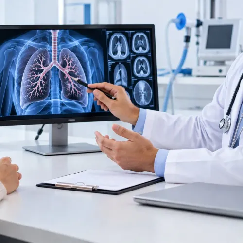
A PET-CT maps the primary tumour and searches for spread, but an avid spot is not automatically cancer. Lung-cancer staging combines TNM findings with tissue confirmation, especially when treatment would change.
- PET-CT identifies suspicious lymph nodes and distant sites; infection and tuberculosis can also appear active.
- EBUS-TBNA samples mediastinal lymph nodes and hilar nodes through the airways. A negative sample does not always exclude cancer when imaging suspicion remains high, so further sampling may be needed.
- Brain MRI, or contrast-enhanced brain CT when MRI is unsuitable, checks for metastases that can alter stage and treatment.
A multidisciplinary team reviews the pathology, molecular results, scans, lung function, general health and treatment goals before recommending a plan. For non-small-cell lung cancer, an operable early-stage tumour may receive lobectomy, segmentectomy or wedge resection with lymph-node assessment. Radiotherapy can replace surgery when surgery is unsafe or target a defined tumour.
- Surgery: Curative-intent option for selected medically operable disease.
- Radiotherapy: Definitive treatment for some localised cancers, or targeted treatment for symptoms and limited spread.
- Systemic treatment: Chemotherapy, immunotherapy or targeted therapy for cancers needing whole-body treatment.
- Combined treatment: Chemotherapy with radiotherapy, or drug treatment before or after surgery, when nodal involvement or tumour biology warrants it.
Small-cell lung cancer usually begins with systemic treatment; limited-stage disease often adds thoracic radiotherapy, while surgery is reserved for rare very early cases.
Preparing for surgery and managing respiratory needs during treatment
A normal oxygen saturation does not prove that lung surgery is safe. The decision depends on the planned resection, cancer stage, general fitness and how much functioning lung will remain.
Preoperative assessment commonly includes:
- Spirometry to measure FEV1 and airflow obstruction.
- Diffusion capacity (DLCO) to show how effectively the lungs transfer oxygen into the blood.
- Predicted postoperative FEV1 and predicted postoperative DLCO, calculated for the lung tissue that will remain.
- Stair-climbing, shuttle-walk or cardiopulmonary exercise testing when lung-function results are borderline.
Low predicted postoperative values increase risk but do not automatically rule out surgery. A thoracic team weighs those results against the operation required. Surgery is mainly used for medically operable early-stage non-small-cell lung cancer; small-cell lung cancer usually starts with systemic treatment, with surgery reserved for a small minority of very early cases.
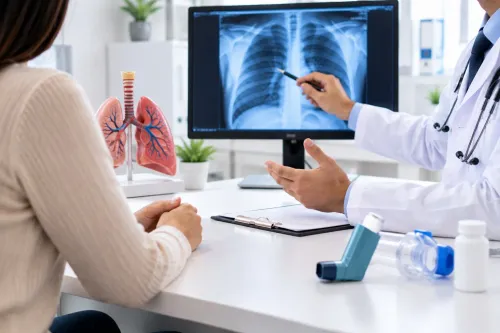
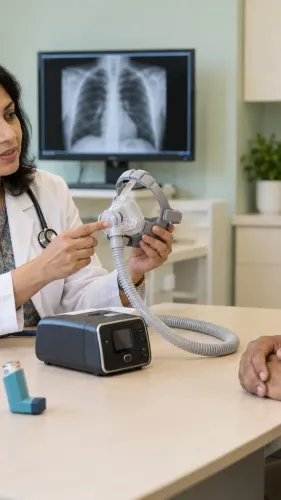
Respiratory treatment alongside cancer care depends on the problem:
- Long-term oxygen therapy requires documented chronic hypoxaemia, such as stable PaO2 of 55 mmHg or lower or saturation of 88% or lower.
- Non-invasive ventilation treats selected acute respiratory failure, including COPD exacerbation with acidosis; confusion, shock or inability to protect the airway may require invasive ventilation and intensive respiratory care.
- Palliative care can begin at diagnosis to control breathlessness, pain, cough, fatigue and caregiver strain while anticancer treatment continues.
What to expect from a coordinated pulmo-oncology service in Trichy
Choose one local clinician or coordinator to keep the pathway together. That person can arrange the pulmonology consultation, retrieve scan images and pathology slides, schedule staging tests, and record the agreed treatment plan so you are not carrying separate reports between departments.
Ask for these items at each review:
- A written diagnosis stating whether the cancer is small-cell or non-small-cell lung cancer
- The stage, biopsy site, and whether lymph nodes or distant organs are involved
- Biomarker results for EGFR, ALK, ROS1, BRAF, KRAS, MET, RET, NTRK and PD-L1 when relevant
- The treatment goal, expected timing, major risks and the next decision date
- A named contact for breathlessness, fever, bleeding or treatment complications
Galaxy Chest & Multi Specialty Hospital Pvt Ltd can be useful when the same local service needs to connect lung-lesion biopsy planning with thoracic surgery and respiratory review. Ask whether your tissue sample is being conserved for molecular testing before treatment begins; a small or exhausted biopsy can delay targeted-treatment decisions.
Keep one folder containing reports, scan discs, medication lists and oxygen readings. Schedule respiratory follow-up during treatment, not only after it, to adjust inhalers, oxygen or ventilatory support. Request palliative care early for breathlessness, pain, cough, fatigue, nutrition or caregiver strain; it can continue alongside anticancer treatment.
Frequently asked questions
When do lung symptoms need urgent assessment or emergency care?
Coughing blood, severe or worsening breathlessness, chest pain, confusion, blue lips or rapidly deteriorating symptoms require emergency care. Persistent cough, unexplained weight loss, hoarseness or a chest infection that does not resolve warrants prompt lung-cancer evaluation.
Can a chest scan confirm lung cancer?
No. A chest X-ray or CT scan can identify a suspicious nodule, mass, enlarged lymph node or spread, but tissue pathology usually confirms cancer and identifies its type.
How is a lung biopsy chosen?
The team selects the safest route based on the lesion’s size and location, lymph-node involvement, lung function and the need for molecular testing. Options include bronchoscopy, endobronchial ultrasound, CT-guided needle biopsy or surgical biopsy.
How do staging and multidisciplinary review affect treatment?
Staging combines imaging, biopsy findings, lymph-node assessment and tests for spread. A multidisciplinary team then matches surgery, radiotherapy, chemotherapy, targeted therapy or immunotherapy to the cancer stage, biomarkers and your respiratory fitness.
How can a pulmo-oncology service support respiratory care during treatment?
Coordinated care can combine cancer treatment with lung-function assessment, bronchoscopy, airway management, infection evaluation, oxygen planning, rehabilitation and perioperative respiratory support when needed.