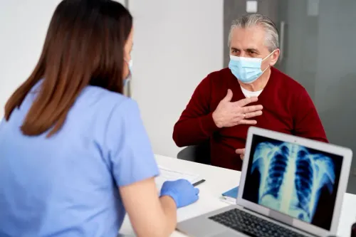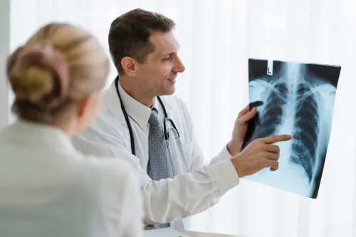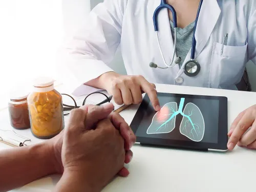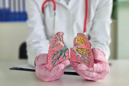

Being told that a chest scan contains a lung nodule, mass or suspicious abnormality can be frightening. However, an abnormal scan does not automatically mean cancer.
Inflammation, infection, old scarring and several non-cancerous conditions can also create abnormalities on chest imaging.
The next step should therefore be careful evaluation by an appropriate lung cancer specialist or multidisciplinary respiratory team rather than assuming the diagnosis before it has been established.
Start With the Imaging Report
The appearance of a lung abnormality helps determine what should happen next.
Doctors may consider characteristics such as:
- Size
- Shape
- Location
- Borders
- Changes compared with older scans
- Presence of enlarged lymph nodes
- Other abnormalities within the lungs
Previous imaging is particularly valuable.
A finding that has remained unchanged for a long period may have different implications from one that is newly appearing or increasing in size.
Patients should therefore carry any older chest X-rays or CT scans to the consultation whenever possible.
Understand That Diagnosis May Require Tissue
Imaging can raise or lower suspicion, but definitive diagnosis often requires examining cells or tissue.
Depending on where the abnormality is located, doctors may consider different methods of obtaining a sample.
These can include bronchoscopy and image-guided biopsy techniques, among other approaches.
The safest and most useful method depends on the individual case.
A specialist should be able to explain why a particular diagnostic approach is being considered.
Ask About Bronchoscopy Capability
Some lung abnormalities are located within or close to the airways.
Bronchoscopy allows a respiratory specialist to examine the airways using a flexible instrument and, where appropriate, obtain samples.
It can also be useful when investigating certain unexplained respiratory symptoms or abnormal imaging findings.
Not every lung nodule requires bronchoscopy, so the procedure should be selected according to clinical need.
Multidisciplinary Care Can Become Important
If lung cancer is confirmed, treatment planning may involve several specialties.
Depending on the type and stage of cancer, care can involve pulmonology, medical oncology, radiation oncology, surgery, radiology and pathology.
This makes coordination important.
A respiratory specialist may initially help establish the diagnosis and assess lung function before the patient proceeds to the relevant cancer treatment team.
Know Which Symptoms Need Prompt Attention
A suspicious scan may sometimes be discovered before significant symptoms appear.
When symptoms are present, they may include:
- Persistent cough
- Blood in the sputum
- Unexplained weight loss
- Chest discomfort
- Increasing breathlessness
- Recurrent infection in the same area
- Persistent hoarseness
These symptoms have many possible causes, but they should not be ignored.
What to Bring to the Appointment
Patients in Trichy attending evaluation for an abnormal lung scan can prepare by carrying:
- The latest CT or chest X-ray images and reports.
- Previous chest imaging for comparison.
- Pathology reports if a biopsy has already been performed.
- Details of previous respiratory illnesses.
- Current medication information.
- A brief smoking and occupational exposure history.
Having these records available can reduce unnecessary duplication and help specialists understand the timeline.
Galaxy Chest & Multi Specialty Hospital Pvt Ltd provides hospital-based respiratory evaluation in Trichy for patients with lung and chest abnormalities, including cases requiring pulmonary investigations.
Choosing a specialist after an abnormal scan should focus on diagnostic readiness. Look for access to respiratory expertise, imaging review, appropriate pulmonary procedures and coordination with other specialties when required.
The immediate goal is not to assume the worst. It is to establish exactly what the abnormality represents and determine the correct next step.